Bimonthly assessment for the month of February

Bimonthly assessment for the month of feb 2021


Submission of bimonthly assessment for the month of feb 2021

Q.1) Please go through the patient data in the links below and answer the following questions:

50 year man, he presented with the c/o
Frequently walking into objects along with frequent falls since 1.5 years
Drooping of eyelids since 1.5 years
Involuntary movements of hands since 1.5 years 
Talking to self since 1.5 years 

More-https://archanareddy07.blogspot.com/2021/02/50m-with-parkinsonism.html?m=1
Case presentation links: https://youtu.be/kMrD662wRIQ

Problem presentation-
1.Involuntary movements of both upper limbs 
2.Drooping of eyelids
3.Talking to self 
4.Frequent falls (while walking on steps) 
All the above from 1.5 years. 
Localisation of lesion-
➡Drooping of eyelids  is called as ptosis
and our patient has Bilateral ptosis
B/l ptosis - is because of weakness in levator palpebrae superioris muscle and muller muscle which is because of ------
1)Muscle involvement 
2) Nerve involvement (3rd cranial nerve) 
3) Nucleus involvement (ie central caudal nucleus of oculomotor complex
https://www.sciencedirect.com/topics/medicine-and-dentistry/levator-palpebrae-superioris-muscle
➡Involuntary movements of upper limbs-we can categorize under movement disorders 

Problem representation and anatomical localization : 

➡️Running into the objects ,drooping of eyelids --EYELID OPENING APRAXIA —

Possible causes and anatomical localization include - 

Muscles involved in opening eyelids 

➡️levator palpebrae superioris-LPS is supplied by occulomotor nerve 

➡️Mullers muscle 

➡️Frontalis muscle


cerebral cortex, basal ganglia, and superior colliculus (SC) are thought to play a role in disinhibition of leavator palpebra superioris 


➡️Orbicularis oculi -closes eyelids 

➡️Cerebral cortex -provides promotor control for eyelid movements,medial frontal lobes including supplementary area involved in control of blinking 

➡️Substabtia Nigra pars reticulata-dopamine depletion decreases blink rate -SEEN WITH PARKINSON PATIENT .

➡️superior colliculus 


EYELID DISORDERS - Droopy eyelids can be caused by neurogenic -innervational failure or LPS damage -myogenic Ptosis 


Causes of neurogenic ptosis include :


third cranial nerve palsy 

➡️Horner syndrome 

➡️Myasthenia gravies 

  ➡️botulism

 ➡️ Gullian Barre syndrome

 ➡️chronic inflammatory demyelinating polyneuropathy 


MYOGENIC PTOSIS :

➡️Congenital ptosis 

➡️Eyelid trauma /surgery 

➡️Mitochondrial myopathy 

➡️Oculi pharyngeal dystrophy 

➡️Myotonic dystrophy 

➡️Herpes zoster 

➡️Chronic topical steroid use 


History of frequent falls :

Falls in elderly patient with background history of slowness and decreased expressiveness - would suggest autonomic instability or postural instability seen with  parkinson patient . 

Cause of falls : 

  ➡️Ataxial rigidity 

  ➡️Bradykinesia 

  ➡️  Loss of postural reflexes 

  ➡️Freezing 

  ➡️  Visual  vestibular component 

B) What is the etiology of the current problem and how would you as a member of the treating team arrive at a diagnosis? Please chart out the sequence of events timeline between the manifestations of each of his problems and current outcomes. 

B/l ptosis-
https://www.medicaleducationleeds.com/paces/ptosis/#:~:text=Differential%20Diagnosis%20of%20ptosis%3A,Horner's%20syndrome

Classification of ptosis-
1.Neuropathic--3rd nerve palsy, horner syndrome
2.Neuromuscular junction  -Myasthenia gravis, Botulism 
3Mechanical-lid tumors 
4.myogenic-myotonic dystrophy 
Arriving at a diagnosis 

The size of pupils o/e normal:rules out horner's or 3rd nerve palsy(as a single nucleus supllies both levator palpebral superioris ,its lesion causing b/l ptosis
myasthenia gravis -
no history of fatiguable ptosis (can be diagnosed by ice pack test

Self talk -frontal lobe lesion 

Frequent falls while climbing steps -indicate vertical gaze of patient affected. 


c)What is the efficacy of each of the drugs listed in his current treatment plan

Treatment : 

1 . Tab. SYNDOPA 110mg PO TID

2.  Tab. METFORMIN 500mg PO OD


Metformin MOA - 

➡️Decreases hepatic gluconeogenesis 

➡️Increases peripheral uptake of glucose

➡️Increases insulin sensitivity 


2) Patient was apparently asymptomatic 2 years back then he developed weakness in the right upper and lower limb, loss of speech.

More here: https://ashfaqtaj098.blogspot.com/2021/02/60-year-old-male-patient-with-hrref.html?m=1

a). What is the problem representation of this patient and what is the anatomical localization for his current problem based on the clinical findings?

problem representation:
➡️Sob on exertion grade 2 progressed to grade 4 ( on rest) with orthopnea and paroxysmal nocturnal dyspnea. 
➡️Bilateral pedal edema up to knee since 2 months
➡️Decreased urine output and generalized weakness since 2months
➡️loss of speech persisted from 2 years after patient had weakness in right upper and lower limb (hemiparesis) 

anatomical localization :
➡️  History suggests sob 2 to 4 (even on rest), orthopnea and paroxysmal nocturnal dyspnea. 

A) systolic heart failure 

B)diastolic heart failure 

His examination findings were Visible apical impulse,  Pericardial bulge, visible pulsations, dilated veinsshift of apex beat to 6th ICS, Thrill at the apex, Loud S1 present, loud P2 present, S3 Accentuating on inspiration- RVS3, Expiration - LVS3

His Ecg shows poor R wave progression

Chest Xray PA shows Cardiomegaly 

His 2Echo is suggestive of Heart failure  DCMP with Hypokinesia at RCA, LCX


Anatomical diagnosis:

The location to his problems is at the Heart, secondary to atherosclerosis of the vessels


Risk factors:

Alcohol

Age of 60 years

Male gender


'In vivo imaging techniques applied in humans and the success of antithrombotic and fibrinolytic therapy in ACS established in practice the role of thrombosis in their pathogenesis. A number of microanatomic mechanisms underlie acute coronary thrombosis. According to autopsy studies—clearly biased toward fatal outcomes—a through-and-through rupture of the plaque’s protective fibrous cap most commonly causes lethal coronary thrombosis. Other mechanisms that account for a minority of fatal coronary thromboses include superficial erosion, intraplaque hemorrhage, and the erosion of a calcified nodule. Thus, physical disruption of the atherosclerotic plaque accounts for almost all acute coronary thromboses.'


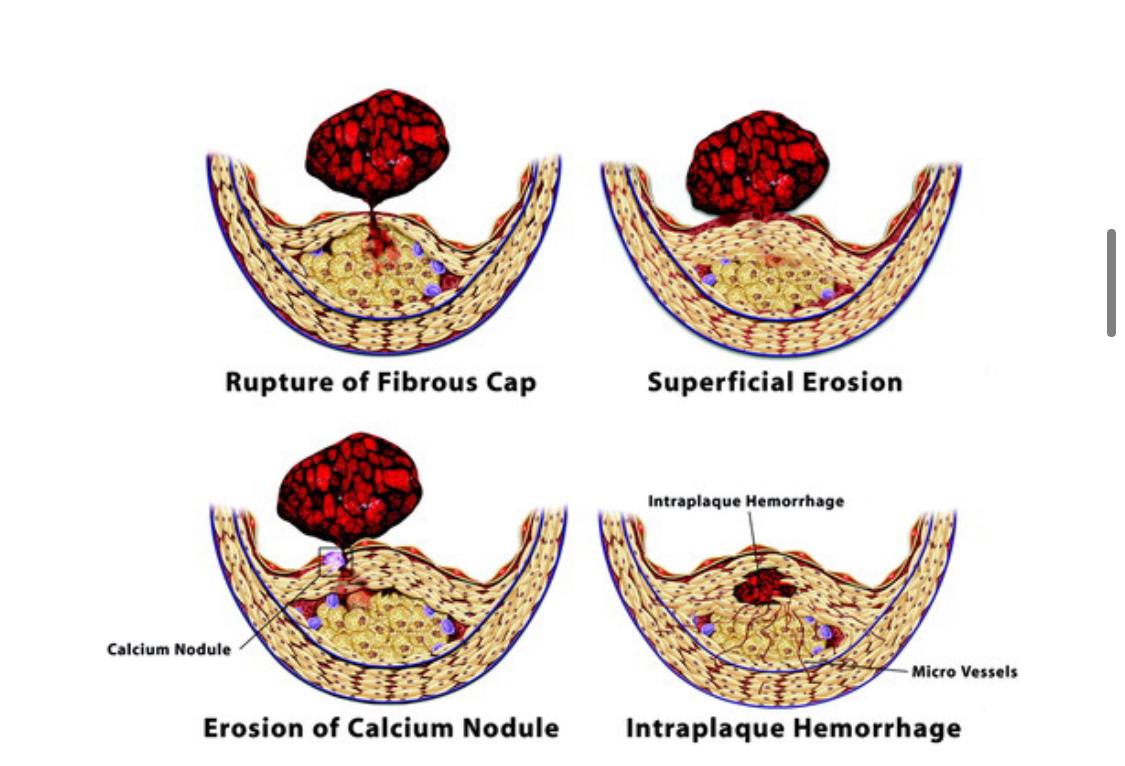
'Disrupted plaques provoke thrombosis in several ways. First, contact with collagen in the plaque’s extracellular matrix can trigger platelet activation. Second, TF produced by macrophages and SMCs activates the coagulation cascade. The disrupted plaque thereby represents a “solid-state” stimulus to both thrombosis and coagulation; these pathways reinforce each other, as thrombin generation amplifies the activation of platelets and other cells in the lesion. Conversion of fibrinogen to fibrin and release of von Willebrand factor from activated platelets can provide the cross-linking molecular bridges between platelets that yield the dense, 3-dimensional network of platelets entrapped in fibrin characteristic of the “white” arterial thrombus.'


Examination findings -
➡️Apex beat at 6th ICS - probably because of ventricular hypertrophy apex beat being displaced. 
➡️Pedal edema upto knee Grade 2 ---maybe because the patient might have developed features of right heart failure too (2nd most common cause) 
➡️Loud p2
➡️Bilateral  fine Crepitations  present in axillary,infra axillary and infrascapular areas.


What is the etiology of the current problem and how would you as a member of the treating team arrive at a diagnosis? Please chart out the sequence of events timeline between the manifestations of each of his problems and current outcomes. 


Etiologies -  1.coronary heart disease -ICMP

                   2.Cardiomyopathies -DCMP

                   3. Valvular heart disease - predominantly left side 

Ecg showing 
1)normal axis
2)pathological Q waves from v1 to v6
3)poor R wave progression
suggest a CAD probably involving LAD and LCX 
sequence of events(probable hypothesis) 
Based on history -male and being addicted to alcohol (quantity and duration not being mentioned) 
               šŸ”½⬇️
Leads to depletion of NADPH and  thus  affects glutathione perioxidase 
               ⬇️
Resulting in formation of ROS reactive oxygen species ➡damages tissues 
 2) Alcohol overintake 
              ⬇️
     Deranged lipid profile 
              ⬇️
     Atherosclerosis of vessels 
              ⬇️
     Affecting coronary vessels 
              ⬇️
   Myocardial infarction
Alcohol metabolism---


 Further events 
Alcoholic cardiomyopathy
      ⬇️
Myocardial infarction
      ⬇️
Ventricular remodelling 
      ⬇️
Ventricular dilation 
Source --https://www.ncbi.nlm.nih.gov/pmc/articles/PMC2365733/

c) What is the efficacy of each of the drugs listed in his current treatment plan 

With no revelant history of smoking and also no wheeze,no seasonal variations -respiratory cause of sob has been ruled out .


Sequence of events :

2 years back weakness of right upper and lower limb with brocas aphasia   

                   F/b 

2 months history of SO,pedal edema 


  1. What is the efficacy of each of the drugs listed in his current treatment plan 


1.Tab. Lasix 40 mg bd


2.Tab. Benfomet plus od


3.Tab. Telma 40 mg od


4.Tab. Met xl 50mg od


5.Tab. Aldactone 25mg od


6.Salt restriction <2gm/day


7.Fluid restriction<1.5lit/day 


Lasix ,fluid and salt restriction are supportive management to decrease preload on the heart .


Spirinolactone + Telmisartan + beta blockers are now recommended therapy for the patient’s with -chronic heart failure with reduced ejection fraction according to recent guidelines . 


Most efficacious is ARNI + other combination .


3)52 year old male , shopkeeper by profession complains of SOB, cough ,decrease sleep and appetite since 10 days and developed severe hyponatremia soon after admission. 

More here https://soumya9814.blogspot.com/2021/01/this-is-online-e-log-book-to-discuss.html?m=1
Case presentation video:
https://youtu.be/40OoVEQBgS4


a) What is the problem representation of this patient and what is the anatomical localization for his current problem based on the clinical findings?


Problem presentation --
➡️
Sob grade 2 progressed to grade 3 
➡️
cough with sputum,whitish in colour non foul smelling since 2 days
➡️
decreased sleep  and appetite since 10 days

Anatomical localisation--
Sob grade 2 to 3 without complains of palpitations , chest pain ,orthopnea and pnd
can be attributed to lung problem. 

b) What is the etiology of the current problem and how would you as a member of the treating team arrive at a diagnosis? Please chart out the sequence of events timeline between the manifestations of each of his problems and current outcomes? 

Ans--
Etiology- 
Based on history -- 
Sob 2 to 3,generalized weakness, decreased appetite (anemia causing heart failure) 
O/E - on examination of lower palpbreal conjuctiva -pallor+

Sequence of events 
50 year old with gradual reduction in hemoglobin 
⬇️
Developed symptoms of sob, ⬇sleep and appetite, generalized weakness 
⬇️
Eventually patient developed heart failure. 
Also he took statins for an year and is a k/c/o type 2 dm 

Approach to this patient : 

We will screen for CAD - as patient falls under high risk for CAD 

General -pallor + 

Systemic examination - no obvious findings of pneumonia 

ECG and echo showed no evidence of CAD ,

Concentric LVH with EF -62 percent .

X ray - bilateral pleural effusion with no consolidators changes .


Hemogram showed dimorphism anemia ( mostly due to Iron and vitB12 deficiency - Nutritional deficiency ) 


Etiology - Dimorphic anemia secondary to iron and vit B12 deficiency 


Patient after admission suddenly developed hyponatremia .

Approach to hyponatremia 


1) First -restricting fluids with minimal intake of hypertonic saline 3%
Calculation of the sodium deficit:
0.6 x weight(kg) x (desired Na+ - Actual Na+). Use 0.5 for females. Desired Na+= 120-125 meq/l.
Example: 70kg male. Na+= 110 meq/l Desired target= 125 meq/l.
[0.6 x 70kg x (125-110)= 630 meq of Na+ needed].
Amount needed to increase serum sodium level by 1 meq/l/hr= 0.6 x 70kg x 1.0= 42 meq/hr (safe rate for this patient).

3%--hypertonic saline contains 513 meq/liter.

Therefore: [desired rate per hr] / 513 x 1000 = infusion rate (ml/hr).
And the total infusion time= [total meq needed] / [meq/hr]
Desired rate= 42/513 meq x 1000= 82 ml/hr
Infusion time= [630 meq] / [42 meq/hr] = 15 hrs.
Therefore: Infuse 3% saline at 82 ml/hr for 15 hours.

Source---
 https://globalrph.com/dilution/sodium-chloride/

➡️Maximum correction for Na which can corrected in a day is 8-10 meq
➡️First 4 hrs -4meq has to be corrected 
➡️Next 20 hrs -remaining calcuated Na should be given 
➡️we have to control sugars since for every 100mg increase in sugars, 1.6 Na will decrease .

However in a type 2 diabetic we consider 124 too as normal Na range 
Source of the above points =2-4 Class 

2) proper control of sugars with insulin and metformin 


c) What is the efficacy of each of the drugs listed in his current treatment plan especially for his hyponatremia? What is the efficacy of Vaptans over placebo? Can one give both 3% sodium as well as vaptan  the same pt? 


Answer--
1) Efficacy of vaptans over placebo
https://acsjournals.onlinelibrary.wiley.com/doi/full/10.1002/cncr.28468
2) Can one give both 3% sodium as well as vaptan to the same patient?  
No, 
We shouldn't give both at a time. 
https://www.ncbi.nlm.nih.gov/pmc/articles/PMC5752787/

Q.4 .Please mention your individual learning experiences from this month.

1.Understood relations, medical ethics, medical etiquette. 
2.Calculation of the sodium deficit:
0.6 x weight(kg) x (desired Na+ - Actual Na+). Use 0.5 for females. Desired Na+= 120-125 meq/l.
Example: 70kg male. Na+= 110 meq/l Desired target= 125 meq/l.
[0.6 x 70kg x (125-110)= 630 meq of Na+ needed].
3.Barrel shaped chest4.Engorged veins5.Angular stomatitis seen in chronic anemic patient-PLUMMER VILSON SYNDROME

Popular posts from this blog

70 yrs M with CAD wirh HFrEF with( EF 35%) LAD with AF with FVR with LBBB

42/ M WITH HFPEF WITH ALCOHOLIC DEOENDANCE SYNDROME WITH CHRONIC LIVER DISEASE.

chronic decompensated liver disease secondary to alcohlolism